Treating Different Types of Cracks in Teeth
Cracks are one of the most common dental pathologies after caries and periodontal disease, yet many practitioners lack confidence in their approach to crack diagnosis and treatment. Traditional recommendations for treating cracked tooth syndrome are to crown the tooth and, if symptoms persist, perform endodontic treatment.
Drawing on research from the last two decades, we now have a deeper understanding of crack initiation in teeth, how cracks present symptoms and best practices for treatment to conserve tooth structure and preserve pulp vitality long-term.
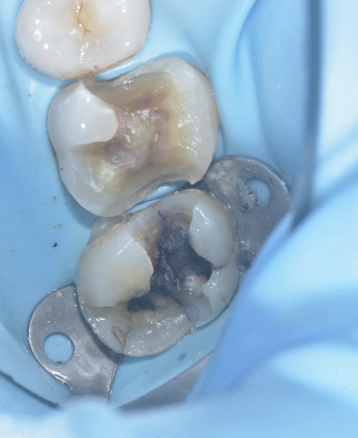
Using Dr. David Alleman, DDS’s Six Lessons Approach to Biomimetic Dentistry, teeth with deep cracks, like this case by Dr. Davey Alleman, DMD, can be treated predictably while protecting the pulp and critical tooth structure.
What causes cracks in teeth?
Crack initiation is a topic well-known to engineers but not for dentists. Cracks form when a material fatigues over time from bending and flexing. Natural teeth have resilient dentin surrounded by strong enamel, making them resistant to cracks. Cracks into enamel are a tooth’s natural defense against long-term use, but when these cracks extend past the dentin-enamel junction, they compromise the tooth's structure and should be treated like a pathology. While some virgin teeth will crack over time, most cracks initiate after a tooth is restored.
Sub-occlusal transverse ridges in posterior teeth, also known as Rainey Ridges after researcher Tim Rainey, support the top of the tooth under the forces of occlusion. When these are removed during a large cavity or crown preparation, and the restoration is poorly bonded or relies on retention form, the tooth bends and flexes more than it is naturally designed to move. This can lead to crack formation around or underneath the restoration.
Learn more about crown alternatives for cracked teeth.
The traditional preparations in these teeth have removed the sub-occlusal transverse ridges on these teeth, making them more prone to cracks. Once the restorations are removed, cracks are visibly extended through the enamel and into the dentin.
Case by Dr. Davey Alleman, DMD
Types of cracks in teeth
There are three main types of cracks into dentin that should be of concern to practitioners.
Peripheral rim fracture: Common around the outsides of restorations, peripheral rim fractures are often first diagnosed as poor dental hygiene that has resulted in a cavity. Look for symmetrical caries around the crack (this is most easily done with caries detector dye) to determine if the crack is the cause of the lesion.
Horizontal/oblique cracks: These cracks are often found around a restoration that is surrounded by thin cusps. Without the support of the sub-occlusal transverse ridges, these cusps can crack over time from the forces of occlusion.
Vertical cracks: Vertical cracks are the most dangerous because of their proximity to the pulp. Typically found under restorations, these cracks are also the most difficult to treat and visualize.
Peripheral rim fractures extending into the dentin are visible in these teeth that were previously restored with traditional preparations. Case by Dr. Davey Alleman, DMD.
A horizontal/oblique crack was responsible for this tooth’s fractured cusp in this case by Dr. Davey Alleman, DMD. The previous composite was not bonded to the tooth to adequately support the remaining tooth structure during the forces of occlusion
Vertical cracks extending across the tooth and near the pulp are visible in this case by Dr. Davey Alleman, DMD.
Understanding the different types of cracks into dentin while aid in diagnosis and treatment. The Six Lessons Approach gives doctors a 1-2-3-4 risk assessment for diagnosing teeth that are at risk of cracking due to structural compromise. Additional research into dentinal pain and how cracks grow, supports crack diagnosis based on patient symptom history.
Treatment recommendations for all cracks are the same. A natural tooth is free from gaps in dentin, so restorations without gaps will have the best long-term outcomes. Complete removal within the peripheral seal zone will conserve tooth structure and vitality. This can be done safely, even in deep areas, by following the Six Lessons Approach Lesson 2 protocols. To learn more watch our Free Biomimetic Dentistry Webinar about Crack Management.
The risks cracks pose to tooth health
Like all dental pathologies, the sooner cracks are treated, the better. Cracks can cause pain for patients, and if cracks are left untreated, they will continue to grow. Cracks are a pathway for bacteria to enter the tooth. In the case of a vertical crack, this means a direct line for bacteria to enter near the pulp. Large cracks can lead to loss of tooth structure, whether a cusp from a horizontal/oblique crack or the tooth itself in the case of a vertical crack approaching the root.
Just like caries, early diagnosis and treatment of cracks can help conserve critical tooth structure and protect tooth vitality. Case by Dr. Davey Alleman, DMD.
Biomimetic treatment of cracks
Biomimetic means to mimic nature, so in the case of biomimetic dentistry, our aim is to mimic a natural tooth. A natural tooth is free of gaps or cracks into dentin around the pulp chamber, so a tooth restored biomimetically should be too. At the Alleman Center, doctors learn steps for safe, predictable crack removal aided by high magnification and photography. Once the crack is removed, the tooth is restored using advanced adhesive techniques like immediate dentin sealing that continue to mimic a natural tooth, bonding to the healthy tooth structure at the same strength at which a natural tooth is bonded to itself. This returns the tooth to natural function, even if the tooth’s sub-occlusal transverse ridges have been removed, making the restored tooth resilient to future crack initiation and free of symptoms.
Drawing on what we know about crack initiation from engineers, covering a crack or bonding over it will not stop the crack from propagating. Occlusal force, whether applied to the top of a crown or composite on top of the crack, will still be transferred to the bottom of the crack, causing it to grow larger. Only by removing the crack can you stop the risk it poses to the tooth and eliminate symptoms.
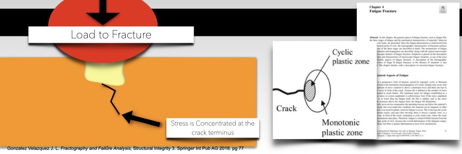
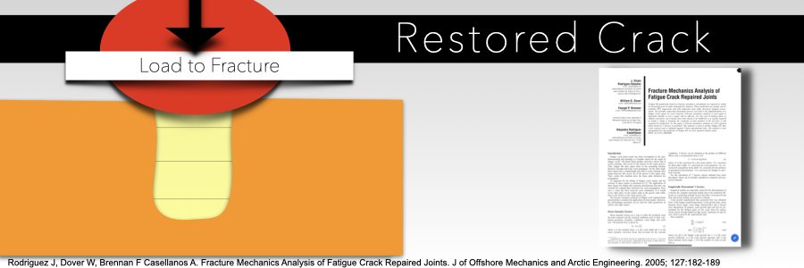
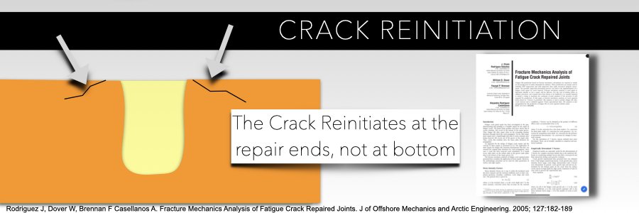
These slides show how a crack that is bonded over will continue to grow when force is applied. Complete crack removal, where possible, protects that deep area of the tooth and transfers the stresses to the top of the tooth, when restored biomimetically.
Alleman Center Biomimetic Mastership slides
Clinical recommendations
The Alleman Center teaches Dr. David Alleman’s Six Lessons Approach to Biomimetic Restorative Dentistry (SLA). This set of protocols gives doctors complete guidance on restoring a tooth in a way that mimics a natural tooth. Lesson 2 of the SLA discusses diagnosis and treatment of cracks while highlighting how a tooth’s natural structure is the guide for reconstruction.
Recommendations for crack diagnosis and treatment include:
Follow the 1-2-3-4 risk assessment that uses patient treatment history and symptoms to aid in the diagnosis of cracks, helping practitioners treat cracks early and more conservatively.
Use 6.5x - 8x magnification or a dental microscope to help visualize cracks. You can’t treat what you can’t see!
Know your limits. The peripheral seal zone concept (Alleman D., Magne P. A systematic approach to deep caries removal endpoints: The peripheral season concept in adhesive dentistry. Quintessence Int. 2012;43(3)197-208.) applies to cracks too. These precise measurements protect the pulp while helping you create a superior bonding field for your restoration.


Here is the full case from Dr. Davey Alleman, DMD. While this treatment took more time than a traditional crown, it prevented endodontic treatment in both teeth, conserved critical tooth structure for natural function and preserved the vitality of these teeth.
When caries is diagnosed during a bi-annual exam, it is treated promptly. Why isn’t this the case for cracks? The answer: traditional techniques and recommendations do not adequately equip doctors to treat cracks in a way that eliminates symptoms and benefits the tooth’s long-term health. However, thanks to advancements in biomimetic dentistry, full-coverage crowns are no longer the only option for crack treatment. Cracks in teeth can now be treated more conservatively, while resolving symptoms and preserving the tooth’s integrity with crown alternatives. To learn more about confident crack treatment, view upcoming Alleman Center training programs.
Published: October 16, 2024
Author: Audrey Alessi
Editor: Dr. David Alleman, DDS
