Cuspal Fracture: Prevention and Treatment
Fractured cusps are a common failure of restored and even virgin teeth. In moderate cases, patients will notice their tooth feels different with their tongue or experience food impaction. In severe cases, patients will experience pain when larger portions of their tooth fracture. In either scenario, these fractures could often have been prevented with better crack diagnosis and restorative protocols, like those in Dr. David Alleman’s Six Lessons Approach to Biomimetic Restorative Dentistry.
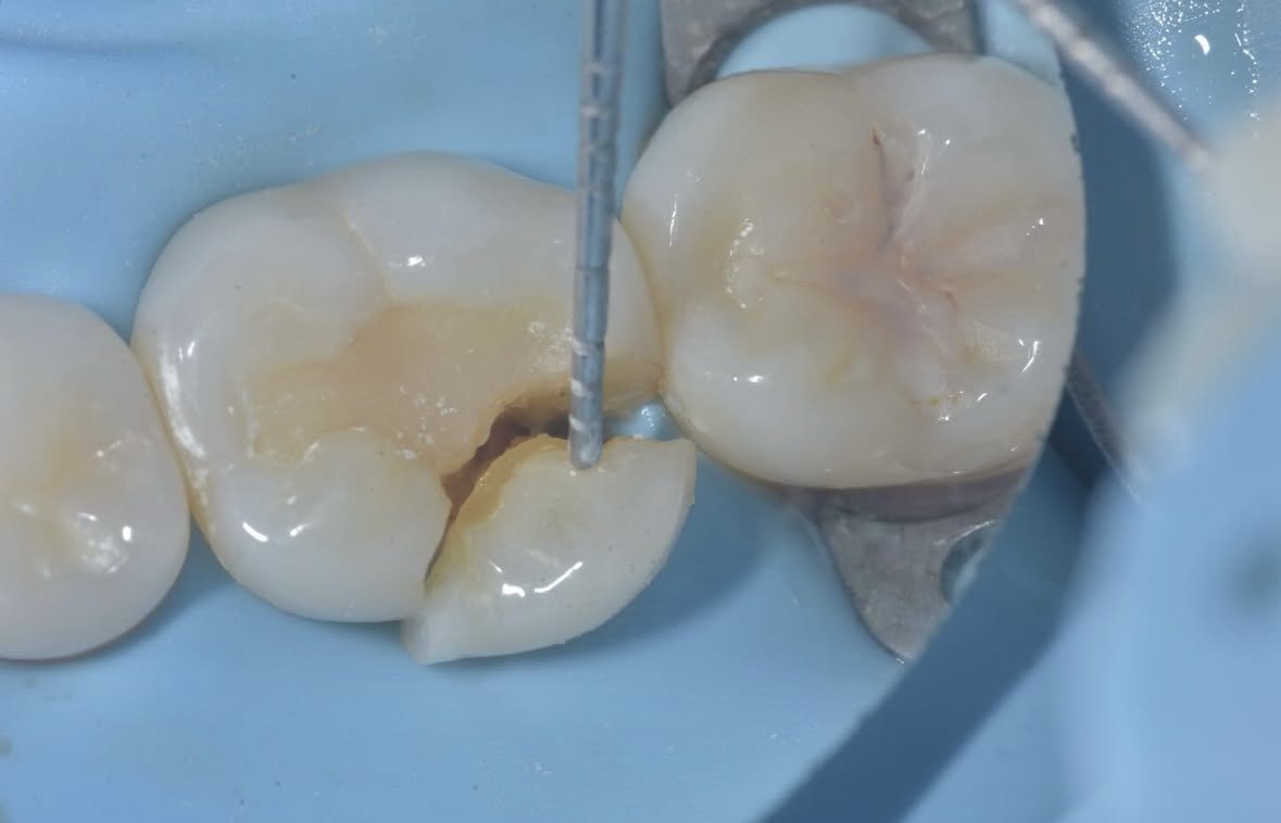
A poorly bonded composite restoration resulted in cuspal fracture as seen in the initial photo in this case by Dr. Davey Alleman, DMD.
Why cusps fracture
Large restorations: large restorations leave thin cusps around the composite or amalgam. These thin cusps often have deep undercuts, reducing their hydration from the dentin tubules, which makes them weaker than fully hydrated cusps. This decreased resilience means less force is required to initiate a crack that would fracture the cusp (Kishen A, Vedantam S. Hydromechanics in dentine: Role of dentinal tubules and hydrostatic pressure on mechanical stress-strain distribution. Dent Materials. 2007; 23: 1296-1306).
The large amalgam restoration left the tooth unsupported, resulting in a cuspal fracture. Case by Dr. Davey Alleman, DMD.
Low bond strengths: retention-form amalgam restorations and poorly bonded composite restorations do not support the remaining structure of the tooth. This means that with every chew, the tooth moves more than it is naturally designed to do (Magne P, Oganesyan T. CT Scan-Based Finite Element Analysis of Premolar Cuspal Deflection Following Operative Procedures. 2009 Quintessence Int, 2009; 29:361-369). This increased movement makes crack initiation more likely, which can result in cuspal fracture.
Just because composite looks like a tooth, does not mean it functions like a tooth. This poorly bonded composite resulted in a cuspal fracture. Case by Dr. Davey Alleman, DMD.
Undiagnosed cracks: undiagnosed cracks in virgin teeth can lead to fractured cusps. Without diagnostic protocols specific to cracks, this pathology can be difficult for dentists to diagnose. If allowed to propagate, horizontal cracks can lead to a fractured cusp. Learn more about early diagnosis of cracks in Dr. David Alleman’s Six Lessons Approach Podcast.
A crack formed in this virgin tooth. Thanks to early diagnosis, the tooth was restored conservatively by Dr. Davey Alleman, DMD.
Preventing cuspal fracture
1-2-3-4 Risk Assessment: Dr. David Alleman, DDS created a 1-2-3-4 risk assessment for structural compromise that offers guidance for doctors on whether or not a tooth is at risk of cracks, including cuspal fracture. This is one of the steps in his Six Lessons Approach to Biomimetic Restorative Dentistry that offers precise protocols for every restorative step, from diagnosis to treatment.
Learn more about the 1-2-3-4 risk assessment in our free Biomimetic Dentistry webinar by Dr. Davey Alleman and Dr. Dafina Doberdoli.
High magnification: Using 6.5x - 8x magnification or a dental microscope will aid in visualization of cracks. This, along with techniques taught in Alleman Center programs, increase the visibility and predictability of crack treatment.
Treatment of fractured cusps
The natural tooth is connected side to side, front to back and top to bottom. For optimal function and health, a restored tooth should too. A crack into dentin is a disconnection of the tooth and should be removed before it grows larger and puts the pulp at risk. A fractured cusp shows clearly how this disconnection within the dentin can be destructive to the tooth, but while the loss of a cusp is not ideal, this is non-critical tooth structure and can be restored to mimic a tooth’s natural form and function. Onlaying a cusp to remove a crack is preferable to leaving a crack only to have it propagate towards the root or the pulp.
With the advanced adhesive principles of biomimetic restorative dentistry, fractured cusps can be restored predictably with a long-lasting bond.
This case by Dr. Davey Alleman, DMD shows how a tooth with a fractured cusp can be treated using biomimetic restorative dentistry.
Biomimetic secure bond
After Lesson 2 of Dr. David Alleman’s Six Lessons Approach to Biomimetic Dentistry, Diagnosis and Treatment of Cracks, Lessons 3 and 4 create a secure bond to the tooth. This functions as a fail-safe for the remaining tooth structure, so any failures, like a fractured cusp, occur on the restorative material rather than the tooth itself. This fail-safe protects the tooth’s vitality and makes any failures easy and non-invasive to restore. To achieve these results in your own office, learn the Six Lessons Approach with Dr. David Alleman and the Alleman Center team in our upcoming dentistry training programs.
Learn more about early diagnosis of cracks in Dr. David Alleman’s Six Lessons Approach Podcast.


